Samani, a small coastal town on the island of Hokkaido, Japan, has made the most of its proximity to the Horoman peridotite body, which includes Mt. Apoi, site of a natural park devoted to "learning about the earth's transformation from peridotite". The town square in Samani (pictured) has been an open-air petrological museum, displaying a wide variety of locally-occurring peridotites.
Unfortunately, Samani experienced a 3.7-meter wave during the tsunami of March 11, 2011. I don't know more than that about the fate of the town, but since the town square was only few tens of meters from the shoreline, I fear the museum may no longer exist. I've e-mailed the town authorities and am awaiting a reply.
This blog provides a selection of images - mostly photomicrographs - of peridotites. Comments and questions are welcome. If you got here via a search engine, check out the blog archive (at right) - There's a lot to see. If you want to enlarge an image beyond what the interface allows, use "save image as", or drag it (the enlarged image) to your desktop and enlarge it further with graphics software or with your browser.
Friday, April 29, 2011
Monday, April 25, 2011
Magnesite in dunite
 |
| Click on the image to enlarge. Photo: Dan Snyder |
Friday, April 22, 2011
Anthophyllite in dunite
 |
| Click on image to enlarge. Photo: Dan Snyder |
Thanks to Dr. Samuel Swanson of the University of Georgia for directing me to this exposure.
Saturday, April 16, 2011
Webster-Addie ultramafic body, NC - chromite and chlorite in dunite.
 |
| Click on the image to enlarge. Photo: Dan Snyder |
Wednesday, April 13, 2011
Webster-Addie ultramafic body - sheared dunite, full thin section
 |
Click on the image to enlarge. Click twice to enlarge more- it's a lot more interesting close up. Photo: Dan Snyder. |
Tectonized, sheared dunite. Upper 2/3 of image is composed mainly of polygonal olivine grains (more saturated colors) with scattered pyroxene grains (grays and pale yellows), especially at top left; lower 1/3 is composed of sheared talc clots with crushed and elongated grains of olivine and pyroxene. By itself, this thin section probably wouldn't fit the conventional definition of dunite - too much pyroxene - but it was part of a more extensive exposure that is clearly dunite. Webster-Addie ultramafic body, Blue Ridge Mountains, Jackson County, western North Carolina. XPL. Imaged area 22 mm x 39 mm.
Sunday, April 10, 2011
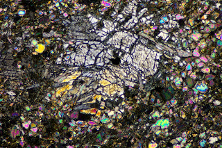
Pyroxene in dunite
 |
| Click on the image to enlarge. Photo: Dan Snyder |
Thursday, April 7, 2011
Olivine weathering microtexture in dunite
 |
| Click on the image to enlarge. Sample:Michael Velbel; Photo: Dan Snyder |
Thanks to Dr. Michael Velbel, Michigan State U., for the loan of this and many other thin sections.
Wednesday, April 6, 2011
Etch pits in olivine - Webster-Addie ultramafic body
 |
| Click on image to enlarge. Photo © Daniel R. Snyder |
The etch pits stand out more clearly in this PPL image of the same feature (same scale as above):
 |
| Click on image to enlarge. Photo © Daniel R. Snyder |
Tuesday, April 5, 2011
Weathering microtexture in olivine.
 |
| Click on the image to enlarge. Sample: Michael Velbel; photo: Dan Snyder |
Thanks to Dr. Michael Velbel, Michigan State U., for the sample, and thanks to Dr. Velbel and NASA for the SEM time. Thanks also to E. Danielewicz for assistance with electron microscopy.
Monday, April 4, 2011
Pyroxene lamellae, amphibole in dunite
 |
| Click on image to enlarge. Photo: Dan Snyder |
Exsolution lamellae in pyroxene (pale yellow grains at left and bottom, gray grain at right); ampohibole mineral (grains with diagonal cleavage patterns: center, top center); olivine (brightly colored grains at center and top center). Webster-Addie utlramafic body, western North Carolina. XPL. Imaged area 1.3 mm x 2 mm.
Thanks to Dr. Michael Velbel, Michigan State U., for the loan of this and many other thin sections.
Subscribe to:
Posts (Atom)
