Friday, June 10, 2011

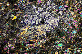
Pyroxene grains and pseudomorphs in Josephine serpentinized peridotite.

Click on image to enlarge.          Photo © Daniel R. Snyder
In this specimen,  A few very small remnants of olivine (bright-colored grains) can be seen around the image, but almost all of the olivine has been altered to serpentine. I collected this specimen on the border between the Josephine peridotite and the Onion Camp complex of Yule et al. (2006)*. I decided to call it Josephine peridotite for two reasons: 1. There is little evidence of shearing, and 2. Many large pyroxene grains still persist, and large serpentine pseudomorphs after pyroxene are distributed throughout the specimen. The large gray pyroxene grain at the left side of the image above still appears to be mostly pyroxene, while the broken grain in the center has been altered to bastite-texture serpentine. Hourglass-texture serpentine, probably an alteration product of olivine, occupies the rest of the image. Klamath Mountains, Josephine County, southwest Oregon, XPL. Imaged area 2.7 mm x 4 mm.

In the image below, the white spots on the large hand specimen are reflections from the surfaces of platy pseudomorphs after pyroxene; gray-green grains are pyroxene. Note that there appears to be more remnant pyroxene than pseudomorphosed pyroxene. The brown mesostatis is serpentine. The surface of the small hand specimen has been rough polished, showing dark grains of pyroxene. Lighter gray areas are serpentinized pyroxene grains (pseudomorphs). Scale is in centimeters.

Click on image to enlarge.          Photo © Daniel R. Snyder

*Yule, J. D., Saleeby, J. B., and Barnes, C. G., 2006, A rift-edge facies of the late Jurassic Rogue-Chetco arc and Josephine ophiolite, Klamath Mountains, Oregon, in Snoke, A. W. and Barnes, C.G., eds., Geological studies in the Klamath Mountain province, Californa and Oregon: A volume in honor of William P. Irwin: Geological Society of America Special Paper 410, p. 53-76.

Monday, June 6, 2011

Peridotite in advanced stage of serpentinization - Onion Camp complex, Oregon

Click on image to enlarge.         Photo © Daniel R. Snyder
The serpentinized peridotite of the Onion camp complex, which is in contact with the variably serpentinized Josephine peridotite, is described by Yule et al. (2006)* as "highly serpentinized and sheared dunite, harzburgite, and sparse websterite....".  In the center of the image above, a pyroxene grain has been pseudomorphosed, probably by an amphibole mineral, with the chain structure preserved as parallel strands of the replacement mineral. A large serpentine pseudomorph after olivine occupies the center of the left side of the image, and a less obvious pseudomorph is at the upper right. Elsewhere in the image, serpentine is ubiquitous. Klamath Mountains, Josephine County, southwest Oregon. XPL. Digital mosaic, imaged area 3.6 mm x 6 mm.

Click on image to enlarge.          Photo © Daniel R. Snyder
Hand samples of serpentinized peridotite of the Onion Camp complex. Smaller sample on right has been rough polished on cut surface. Note hematite coating on left sides of both samples. Scale in centimeters.


*Yule, J. D., Saleeby, J. B., and Barnes, C. G., 2006, A rift-edge facies of the late Jurassic Rogue-Chetco arc and Josephine ophiolite, Klamath Mountains, Oregon, in Snoke, A. W. and Barnes, C.G., eds., Geological studies in the Klamath Mountain province, Californa and Oregon: A volume in honor of William P. Irwin: Geological Society of America Special Paper 410, p. 53-76.

Sunday, May 29, 2011

Second alteration cycle - serpentine altering to anthophyllite (Presque Isle peridotite)

Click on image to enlarge.          Photo © Daniel R. Snyder
The grain shown in this image, originally olivine, was altered to serpentine, most of which has been altered in turn to anthophyllite. That is, I thought it was anthophyllite, but then I read Michael Lewan's (1972)* M. S. thesis, in which he reported NO anthophyllite in the three samples of Presque Isle peridotite for which he carried out modal analysis. However, all three samples contained small amounts of chlorite (7.3, 7.1, and 5.4 percent volume. I've never seen chlorite that looks like this, but maybe... If you know, please send me a comment.

Tiny red or orange anthophyllite(?) crystals occupy most of the former olivine grain, except for the two remaining areas of serpentine, gray-green in the XPL image above (lower left and upper right). The image below, also XPL, shows most of the anthophyllite(?) crystals (those aligned from upper left to lower right, or vice versa) at extinction - as extinct as they get. The serpentine areas show more clearly in this view, while the anthophyllite(?) crystals that are "at extinction" have dark outlines of varying thickness.

Click on image to enlarge.          Photo © Daniel R. Snyder
In the PPL image below, the serpentine areas are relatively featureless, while elsewhere the elongated crystals of anthophyllite(?) show distinct outlines. Yellow-orange color in PPL image is due to iron-oxide staining from hematite. Black spots are magnetite. Presque Isle peridotite, Marquette County, northern Michigan. Imaged area 0.5 mm x 0.8 mm.

Click on image to enlarge.          Photo © Daniel R. Snyder

*Lewan, Michael D., 1972, Metasomatism and Weathering of the Presque Isle Serpentinized Peridotite, Marquette, Michigan, unpublished M.S. thesis, Michigan Technological University.

Presque Isle peridotite - full thin section

Click on image to enlarge.          Photo © Daniel R. Snyder
This image shows the texture of the peridotite, in which olivine and pyroxene have been almost entirely pseudomorphed by serpentine, much of which has been replaced in turn by amphiboles. Light-colored areas are carbonate veins. Marquette County, northern Michigan. XPL Macrophotograph. Imaged area 25 mm x 41 mm.

Friday, May 27, 2011

Plagioclase in Yellow Dog peridotite (plagioclase-bearing lherzolite)

Click on image to enlarge.          Photo © Daniel R. Snyder

 Orthopyroxene at upper left (gray), enclosing plagioclase laths; clinopyroxene (magenta) at lower right; olivine at lower left, upper right, and top center. Yellow Dog Plains, Marquette County, northern Michigan. XPL. Imaged area 2.7 mm x 4 mm (2X objective).

This is an unusually rich concentration of plagioclase (center) for the Yellow Dog peridotite. Most of the plagioclase in my samples is scattered as single laths, perhaps two or three small grains in a typical frame this size.  Older geology texts rule out ANY plagioclase if a rock is to be called "peridotite", but the IUGS classification allows up to 10 percent, in which case they are called "plagioclase-bearing ____". However, the consultant's report states that the plagioclase can average 25 to 30 percent "over significant intervals". In order to accommodate this, the consultant uses a maximum of 30% plagioclase for peridotite. The report defines rocks with between 10% and 30% plagioclase as "feldspathic peridotite".  (This report is on the Web. You can get it in PDF format by Googling "Eagle Deposit Geology".) Under the IUGS classification, these rocks would either be olivine gabbro, olivine norite, or olivine gabbronorite.


Friday, May 20, 2011

Weathered dunite, Newdale, NC

Click on image to enlarge          Photo © Daniel R. Snyder
Above: Weathered dunite, with small grain size and indented grain boundaries resulting from cataclasis and subsequent weathering. All of the blue-green olivine grains clustered at left center go extinct simultaneously, indicatng that they are fragments of a single crystal. Note the dense network of dissolution channels and voids (black). Newdale, Yancey County, North Carolina. XPL digital mosaic. Imaged area 1.3 mm x 3.2 mm.

Below: Optical scan of a freshly-sawn surface of weathered Newdale dunite, same sample as above (NOT same scale). Yellow-orange color is imparted by hematite, disseminated in serpentine layers between and within olivine grains. In the high-magnification view, many of the more robust olivine grains can be seen to retain a light yellowish-green color. Dark grains at lower left are chromite. Dark green grains in cluster at upper left may be remnants of a fragmented hornblende grain. 2400-dpi optical scan. Imaged area 11.4 mm x 17 mm.

Click on image to enlarge.          Photo © Daniel R. Snyder

Talc in weathered Newdale dunite

Click on image to enlarge.          Photo © Daniel R. Snyder
In the above XPL image, a large talc clot (center) is surrounded by weathered olivine grains. The solid black grains at top center, bottom center, and bottom left are chromite. Linear patterns in the lower part of the talc suggest that this part of the talc is partially-altered tremolite/actinolite. Below is a PPL image of the same area at the same scale, showing clearly the chromite and the serpentine meshwork in the dunite. Newdale, Yancey County, western North Carolina. Imaged area 2.7 mm x 4 mm.

Click on image to enlarge.          Photo © Daniel R. Snyder

Wednesday, May 18, 2011

Talc in pyroxene in Webster dunite

Click on image to enlarge.          Photo © Daniel R. Snyder
A vein of talc (lower left to upper right) bisects two large grains of orthopyroxene, surrounded by olivine, in dunite. Webster-Addie ultramafic body, Blue Ridge Mountains, Jackson County, western North Carolina. XPL. Imaged area 2.7 mm x 4 mm.

Tuesday, May 17, 2011

Deformation bands in olivine

Click on image to enlarge.          Photo © Daniel R. Snyder
In this image of dunite, several large olivine grains (center, top left, top right, bottom right) show deformation banding. Webster-Addie ultramafic body, Blue Ridge Mountains, Jackson County, Western north Carolina. XPL. Imaged area 2.7 mm x 4 mm.

Saturday, May 7, 2011

Twin Sisters dunite - cataclasis

Click on image to enlarge.          Photo © Daniel R. Snyder


This image illustrates the cataclastic texture described by Ragan (1963)*. The large subhedral olivine grain is inferred to have formed in the "primary"phase, during which the relatively intact igneous body was emplaced in the crust in the solid state. Subsequent strain resulted in the deformation banding seen in many of the grains, the strain lamellae in the large grain, and the fracturing that produced the mosaic of small grains. Later recrystallization of some small grains caused the strain features to be eliminated, as in the tight cluster at the lower right, where each of the grains shows a uniform interference color. Cascade Mountains, Whatcom County, northwestern Washington. XPL. Imaged area 2.7 mm x 4 mm. Link to Dan McShane's Twin Sisters page.


*D.M. Ragan (1963), Emplacement of the Twin Sisters Dunite, Washington, American Journal of Science, v. 261, p. 549-565.

Twin Sisters dunite - full thin section

Click on image to enlarge.          Photo ©  Daniel R. Snyder
Thin section of Twin Sisters dunite showing strained olivine and pyroxene grains with deformation banding. Cascade Mountains, Whatcom County, northwestern Washington. Macrophotograph, XPL. Imaged area 21 mm x 40 mm. Link to Dan McShane's Twin Sisters page.

Sunday, May 1, 2011

Pseudomorphs after pyroxene in ancient (1.2 Ga) metaperidotite

Click on the image to enlarge.      Sample: Jeff Chiarenzelli; Photo: Dan Snyder

A small body of highly-metamorphosed rocks jn the Adirondack lowlands has been interpreted as metaperidotite because of its CIPW normative mineralogy (harzburgite) and because pseudomorphs after ferromagnesian minerals have been found there (Chiarenzelli et al., 2011)*. The protolith has been dated at ~1.2 Ga using detrital zircons. In this image, two pseudomorphs after pyroxene, now probably consisting of an amphibole mineral, can be seen at left center and top center. Pyrites Complex, St. Lawrence County, northern New York. XPL. Imaged area 1.3 mm x 2 mm.

Thanks to Dr. Jeff Chiarenzelli, of St. Lawrence U., for the loan of this and many other thin sections.

* Jeff Chiarenzelli, Marian Lupulescu, Eric Thern, and Brian Cousens (2011), Tectonic implications of the discovery of a Shawinigan ophiolite (Pyrites Complex) in the Adirondack lowlasnds, Geosphere, 2011;7;333-356

Friday, April 29, 2011

Samani - the town that loves peridotites

Samani, a small coastal town on the island of Hokkaido, Japan, has made the most of its proximity to the Horoman peridotite body, which includes Mt. Apoi, site of a natural park devoted to "learning about the earth's transformation from peridotite". The town square in Samani (pictured) has been an open-air petrological museum, displaying a wide variety of locally-occurring peridotites.

Unfortunately, Samani experienced a 3.7-meter wave during the tsunami of March 11, 2011. I don't know more than that about the fate of the town, but since the town square was only few tens of meters from the shoreline, I fear the museum may no longer exist. I've e-mailed the town authorities and am awaiting a reply.

Monday, April 25, 2011

Magnesite in dunite

Click on the image to enlarge.           Photo: Dan Snyder
A cluster of magnesite (MgCO3) grains in Newdale dunite. Magnesite commonly occurs where peridotites are permeated by carbonate-rich fluids.  Light gray, darker gray, and pale yellow grains at center and left are magnesite. Dark gray grains are at partial extinction; pale yellow grains probably contain a small amount of iron carbonate (siderite) in solid solution. The partially extinct (medium gray) grain at the upper edge of the cluster gives an idea of grain size. Colored grains at bottom, upper left, and right are olivine. Newdale, Yancey County, North Carolina. XPL. Imaged area 2.7 mm x 4 mm. Link to Carolina Geological Society 1997 Guidebook (PDF).

Friday, April 22, 2011

Anthophyllite in dunite

Click on image to enlarge.            Photo: Dan Snyder
Anthophyllite in Newdale dunite. Irregularly shaped grains are olivine; long, straight grains are anthophyllite. Newdale, Yancey County, North Carolina. XPL. Imaged area 1.3 x 2 mm. Link to Carolina Geological Society 1997 Guidebook (PDF)

Thanks to Dr. Samuel Swanson of the University of Georgia for directing me to this exposure.

Saturday, April 16, 2011

Webster-Addie ultramafic body, NC - chromite and chlorite in dunite.

Click on the image to enlarge.           Photo: Dan Snyder
A chromite grain (solid black) in the process of altering to chlorite (gray and gray-brown blades). Surrounding (colored) grains are olivine. Chromite is a ubiquitous accessory in most peridotites of the southern Appalachians. The chlorite mineral is kammererite, a chlorite variety unusually rich in chromium and magnesium. Thus, it grows at the expense of both chromite (FeCr2O4), from which it takes chromium, and olivine, from which it takes magnesium and silica. Webster-Addie ultramafic body, Blue Ridge Mountains, Jackson County, western North Carolina. XPL. Imaged area 1.3 mm x 2 mm.

Wednesday, April 13, 2011

Webster-Addie ultramafic body - sheared dunite, full thin section

Click on the image to enlarge. Click twice to enlarge more- it's a lot more 
interesting close up.      Photo: Dan Snyder.
Tectonized, sheared dunite. Upper 2/3 of image is composed mainly of polygonal olivine grains (more saturated colors) with scattered pyroxene grains (grays and pale yellows), especially at top left; lower 1/3 is composed of sheared talc clots with crushed and elongated grains of olivine and pyroxene. By itself, this thin section probably wouldn't fit the conventional definition of dunite - too much pyroxene - but it was part of a more extensive exposure that is clearly dunite. Webster-Addie ultramafic body, Blue Ridge Mountains, Jackson County, western North Carolina. XPL. Imaged area 22 mm x 39 mm.

Sunday, April 10, 2011

Pyroxene in dunite

Click on the image to enlarge.           Photo: Dan Snyder
A large orthopyroxene grain in dunite. According to the IUGS classification, a dunite is a rock made up of ferromagnesian minerals, mainly olivine and pyroxene, of which more than 90% is olivine (discounting accessory minerals). In practice, serpentine inferred to be derived from olivine is counted as olivine. Although a pyroxene grain dominates this photograph, there are only a few such grains in the sample.  Webster-Addie ultramafic body, Jackson County, North Carolina. Reflected-light photomicrograph of polished block. Ordinary light. Imaged area 3.3 mm x 4 mm.

Thursday, April 7, 2011

Olivine weathering microtexture in dunite

Click on the image to enlarge.      Sample:Michael Velbel; Photo: Dan Snyder
Characteristic diamond-shaped etch pits in olivine. The etch pit field is adjacent to a microfracture in the olivine, through which fluid has permeated, creating the etch pits. Orange areas are iron-oxide mineral stains. Webster-Addie ultramafic body, Jackson County, western North Carolina. Plane-polarized light (PPL). Magnification 400X.

Thanks to Dr. Michael Velbel, Michigan State U., for the loan of this and many other thin sections.

Wednesday, April 6, 2011

Etch pits in olivine - Webster-Addie ultramafic body

Click on image to enlarge.          Photo © Daniel R. Snyder
The dark specks that appear to be mineral grains or polishing grit are actually microscopic etch pits  along fractures in and around this olivine grain. These fractures provided access to fluids that developed weathering textures in the olivine. Webster-Addie ultramafic body, Blue Ridge Mountains, Jackson County, western North Carolina. XPL. Imaged area 0.5 mm x 0.8 mm.

The etch pits stand out more clearly in this PPL image of the same feature (same scale as above):

Click on image to enlarge.          Photo © Daniel R. Snyder


Tuesday, April 5, 2011

Weathering microtexture in olivine.

Click on the image to enlarge. Sample: Michael Velbel; photo: Dan Snyder
Coalescing funnel-shaped etch pits forming serrated edges in olivine (light gray). Darker gray at top and right is serpentine. Webster-Addie ultramafic body, Jackson County, western North Carolina. Scanning electron microscope (SEM) image of polished thin section (backscattered electron image), magnification 650x.

Thanks to Dr. Michael Velbel, Michigan State U., for the sample, and thanks to Dr. Velbel and NASA for the SEM time. Thanks also to E. Danielewicz for assistance with electron microscopy.

Monday, April 4, 2011

Pyroxene lamellae, amphibole in dunite

Click on image to enlarge.           Photo: Dan Snyder

Exsolution lamellae in pyroxene (pale yellow grains at left and bottom, gray grain at right); ampohibole mineral (grains with diagonal cleavage patterns: center, top center); olivine (brightly colored grains at  center and top center). Webster-Addie utlramafic body, western North Carolina. XPL. Imaged area 1.3 mm x 2 mm.

Thanks to Dr. Michael Velbel, Michigan State U., for the loan of this and many other thin sections.

Friday, March 25, 2011

Polyhedral texture in dunite - Scanning Electron Microscope Image

Click on the image to enlarge.  Sample: Michael Velbel; photo: Dan Snyder
Granular (recrystallized) olivine, fractured dunite fragment, Webster-Addie ultramafic body, Jackson County, western North Carolina. Scanning electron microscope (SEM) image (secondary electron image). Magnification 40x.

Thanks to Dr. Michael Velbel, Michigan State U., for the sample, and thanks to Dr. Velbel and NASA for the SEM time. Thanks also to E. Danielewicz for assistance with the SEM.

Tuesday, March 22, 2011

Polyhedral texture (mosaic texture) in dunite

Click on image to enlarge.           Photo © Daniel R. Snyder.
Classic polyhedral mosaic in dunite. This texture results from recrystallization in the solid state, and is common in metamorphic rocks composed mainly of one mineral, such as dunite. Grains tend to have fairly straight sides, and many grain boundaries intersect in triple junctions at approximately 120˚ angles. Webster-Addie ultramafic body, Blue Ridge Mountains, Jackson County, western North Carolina. Macrophotograph, XPL. Imaged area 8 mm x 14 mm.

Sunday, March 20, 2011

Buck Creek ultramafic body, NC - chloritization in dunite

Click on the image to enlarge.           Photo © Daniel R. Snyder
Advanced stage of chloritization in dunite, Buck Creek ultramafic body, Blue Ridge Mountains, Clay County, western North Carolina. Pratt and Lewis (1905)* described the chloritization of the Buck Creek dunite thus: "In some cases the olivine alters extensively to chlorite, which penetrates the grains generally along cleavage cracks first, though frequently in diagonal directions, in long slender laths, sometimes passing uninterruptedly through two or three grains." XPL. Imaged area 1.3 mm x 2 mm.

Thanks to Dr. Brannon Andersen of Furman University for giving me detailed instructions on reaching this exposure.

*Joseph Hyde Pratt and Joseph Volney Lewis, Corundum and the Peridotites of North Carolina.

Saturday, March 19, 2011

Buck Creek ultramafic body - unaltered dunite grading into chloritized dunite

Click on image to enlarge.          Photo © Daniel R. Snyder
Portion of a thin section showing unaltered dunite (left) grading to heavily chloritized dunite (right). Buck Creek ultramafic body, Blue Ridge Mountains, Clay County, western North Carolina. Macrophoto, XPL. Imaged area 10 mm x 20 mm.

Thursday, March 17, 2011

Buck Creek dunite - serpentinized and chloritized


Click on image to enlarge. See image below for enlargement of outlined area.

Polished slab of partially serpentinized and chloritized dunite. Buck Creek ultramafic body, Blue Ridge Mountains, Clay County, western North Carolina. Optical scan at 1524 pixels/cm (600 dpi). Original width 3400 pixels, reduced by Blogger to 1600 pixels at maximum magnification. Slab dimensions 8 cm x 14.5 cm. Below is a higher-resolution image (6096 pixels/cm.) of the 3.5-cm-by-3.5-cm area outlined in the larger image.


Click on image enlarge. Click again to get really close up.

Friday, March 11, 2011

Serpentine meshwork in Josephine peridotite

Click on the image to enlarge.            Photo © Daniel R. Snyder
Josephine peridotite, partially serpentinized. A component of the Josephine ophiolite sequence. Serpentine meshwork occupies space between grains and along fractures within individual grains. Because serpentine can't accommodate the iron given up by the olivine, this is taken up as hematite, which gives the meshwork its brown color. Josephine County, southwest Oregon. XPL Imaged area 1.3 mm x 2 mm.

Tuesday, March 8, 2011

Josephine peridotite - hand samples

Click on the image to enlarge.            Photo © Daniel R. Snyder
Polished section (left) and rough hand sample (right) of Josephine peridotite, a component of the Josephine ophiolite sequence, southwest Oregon and northwest California. Dark grains in polished section (also slightly darker grains in rough hand sample) are orthopyroxene, remainder is olivine. Light areas around edges are weathered rind; white lines are saw marks. Josephine County, Oregon. Scale in centimeters.

Saturday, March 5, 2011

Josephine peridotite (harzburgite) - Full thin section in cross-polarized light

Click on the image to enlarge. Copy the enlarged image to enlarge more - 
It looks much better close up.                      Photo © Daniel R. Snyder
Josephine peridotite, a component of the Josephine ophiolite sequence. Small, brightly colored grains are olivine, larger grains in shades of gray are orthopyroxene.  Josephine County, southwest Oregon. XPL macrophotograph of entire thin section. Imaged area 18 mm x 38 mm.

Wednesday, March 2, 2011

Josephine peridotite - digital mosaic

Ckick on image to enlarge.          Photo © Daniel R. Snyder
In this image: mostly olivine (bright colors) with incipient serpentinization, and a few large (gray) grains of orthopyroxene. Josephine peridotite, a component of the Josephine ophiolite sequence. Klamath Mountains, Josephine County, southwest Oregon. Digital mosaic of four 2x-objective images. XPL. Imaged area 5.1 mm x 7.6 mm.

Saturday, February 26, 2011

Josephine peridotite (harzburgite) - pyroxene

Click on image to enlarge.           Photo © Daniel R. Snyder
Josephine peridotite (partially serpentinized harzburgite), a component of the Josephine ophiolite sequence. Gray grain at left is pyroxene, large gray grain in center is partially serpentinized pyroxene (bastite texture). Brightly colored grains are olivine. Klamath Mountains, Josephine County, southwest Oregon.  XPL. Imaged area 1.3 mm x 2 mm.

Wednesday, February 23, 2011

Serpentinite exposure

Click on the image to enlarge.           Photo © Daniel R. Snyder
Serpentinite exposure adjacent to an extensive occurrence of Josephine peridotite.  Because serpentine can't accommodate the iron in the peridotite protolith, much of this is taken up as hematite, visible as  brick-red layers in the exposure. Road cut opposite Eight Dollar Mountain, Josephine County, SW Oregon. Rock hammer for scale is in the white circle at the bottom of the photograph.

Sunday, February 20, 2011

Josephine Peridotite exposure

Click on the image to enlarge.            Photo © Daniel R. Snyder
Exposure of Josephine peridotite, base of Eight Dollar Mountain, Klamath Mountains, Josephine County, southwest Oregon.

Monday, February 14, 2011

Deer Lake peridotite - magnetite traces of ancient olivine crystals

Click on image to enlarge.          Photo © Daniel R. Snyder
The Deer Lake peridotite, thought to be Proterozoioc in age, is composed entirely of secondary minerals. Here, the outlines of two adjacent olivine crystals - and their internal fractures - have been preserved by stringers of magnetite that separated out when the olivine was serpentinized. The gray masses appear to be carbonate, possibly magnesite, throughout which magnetite grains have been disseminated, giving the gray color. Deer et al.* state that magnesite may be formed if serpentinites undergo low-grade metamorphism where carbon dioxide is available. A few orange and red wisps of anthophyllite or tremolite signal the beginning of another round of alteration. Marquette County, northern Michigan. XPL. Imaged area 1.3 mm x 2 mm.

Deer, W. A., Howie, R. A., and Zussman, J., (1992), An Introduction to the Rock-forming Minerals,  2nd edition, Pearson-Prentis Hall, 685 p.

Sunday, February 13, 2011

Deer Lake peridotite (completely serpentinized)

Cllick on image to enlarge.          Photo © Daniel R. Snyder

The Deer Lake peridotite crops out in a small area north of Ishpeming, in Marquette County, northern Michigan. Although the geologic map of the area labels this exposure as "peridotite" (according to convention), because if its inferred protolith  it is in fact serpentinite. Pictured are two varieties of serpentine, as well as magnetite (black lines and spots) and hematite (reddish-brown areas) from the original olivine and pyroxene, which couldn't be taken up by the serpentine. The magnetite makes this rock weakly magnetic. XPL. Imaged area 1.3 mm x 2 mm. Link to photo of hand samples

Thursday, February 10, 2011

Presque Isle peridotite

Click on the image to enlarge.           Photo © Daniel R. Snyder
Peridotite (thoroughly serpentinized). Pseudomorphs of serpentine after olivine (dark green) and anthophyllite after pyroxenes (linear patterns of brown, orange, and yellow) retain the shapes of original crystals. I've seen the Presque Isle peridotite described as "highly deformed". This doesn't look highly deformed to me. A rock body whose protolith is a billion-plus years old and still preserves the original crystal shapes seems to me to hardly be deformed at all.

Michael Lewan's (1972)* M.S. thesis reports on three samples ranging from 54.9 percent to 59.2 percent serpentine by volume. Olivine plus augite account for another 15.7 to 21.2 percent total, about evenly divided. Lewan also found 4.4 percent to 7.8 percent anorthite in his three samples, but I've looked at several thin sections and haven't seen any. Presque Isle Park, City of Marquette, Marquette County, northern Michigan. XPL. Imaged area 1.3 x 2 mm. Link to photo of outcrop.

*Lewan, Michael D., 1972, Metasomatism and Weathering of the Presque Isle Serpentinized Peridotite, Marquette, Michigan, unpublished M.S. thesis, Michigan Technological University.

Tuesday, February 8, 2011

Tremolite in Yellow Dog peridotite

Click on image to enlarge.         Photo © Daniel R. Snder
Tremolite (center) in Yellow Dog peridotite. Tremolite and actinolite, both calcic clinoamphiboles, are very similiar in appearance, having bladed or fibrous crystals or, as in this image, thin columnar crystals in parallel aggregates. Tremolite is the magnesian end-member of the tremolite-ferro-actinolite solid solution, while iron substitutes for some of the magnesium in actinolite.

Two diagnostic differences are that actinolite is slightly pleochroic and ranges from pale green to deep green in thin section (the greater the iron content, the deeper the green). Conversely, tremolite is colorless in thin section and non-pleochroic. In PPL, this specimen proved to be colorless and non-pleochroic - thus it is tremolite.

Tremolite alters to talc and to chlorite. In the above image, talc borders tremolite on the top and right edges of the bladed portion (granular mineral with very high birefringence), and also forms small clots below it and to the left.  Rounded mineral grains around periphery of image are olivine. Pyroxene (gray) is at right, and mica (red) is at left. XPL. Imaged area 2.7 mm x 4 mm.

Higher magnification image (below) of bladed portion of tremolite bundle and talc border. Gray grain at lower right is serpentine. XPL. Imaged area 1.3 mm x 2 mm.

Click on image to enlarge.          Photo © Daniel R. Snyder


Clinopyroxene altering to tremolite:

Click on image to enlarge.          Photo © Daniel R. Snyder
A large anhedral grain of clinopyroxene (magenta and violet) is undergoing alteration to tremolite (light-colored mineral at left, bottom center, and right). Unlike the tremolite in the example above, the habit in this image is fibrous (asbestiform). The fibers are clearly visible against the background of the dark band of chlorite at the upper right. XPL. Imaged area 1.3 mm x 2 mm.

Marquette County, northern Michigan.

For more information on the Yellow Dog peridotite, see posts from January 28, 2011, and May 27, 2011.  For still more information, scroll to the top of this page and enter "Yellow Dog peridotite" in the search box at the upper right. You can also click on "Yellow Dog peridotite" in the cloud at the bottom of the page. Be sure to look at the last post on a page, and click on "newer posts" or "older posts", since Blogspot doesn't always display everything at once.

Monday, February 7, 2011

Yellow Dog peridotite - chloritization

Click on image to enlarge.          Photo © Daniel R. Snyder
Chlorites are a group of hydrous minerals that occur in igneous rocks, where they are products of the hydrothermal alteration of mafic minerals. Mg-rich clinochlore, Mg5Al2Si3O10(OH)8, is a common alteration product of olivines and pyroxenes. Klasner et al.* reported that chlorite makes up from 5 to 15 percent of the Yellow Dog peridotite. Since olivine and pyroxenes are lacking in aluminum, the aluminum in the chlorite was probably either derived from the small amount of plagioclase in the peridotite, or transported by percolating fluids from the surrounding Michigamme slate, into which the peridotite is intruded.

In this image, a contiguous belt of chlorite (dark blue interference color with light gray patches) runs from upper left to right center. It is irregular in width, with two large areas connected by narrow veins, and extends past the borders of the image. The belt visible in the image is clearly a cross-section of a lumpy layer of chlorite extending beyond the picture plane.  This layer probably follows a former fracture, along which metasomatizing fluids reached the mafic minerals and gradually altered them to chlorite, widening and filling the fracture.

Here, the chlorite is bordered mainly by olivine in the process of alteration. The cores of the olivine crystals are still intact, but the edges adjacent to the chlorite have altered to a white, fibrous mineral, possibly a different form of chlorite, or possibly serpentine, which readily alters to chlorite.

Marquette County, northern Michigan. XPL. Imaged area 2.7 mm x 4 mm.

*John S. Klasner, David. W. Snider, W. F. Cannon, and John F. Slack (1979), The Yellow Dog Peridotite and a Possible Buried Igneous Complex of Lower Keweenawan Age in the Northern Peninsula of Michigan, Geological Survey Division, Michigan Department of Natural Resources, 38 p.

Sunday, February 6, 2011

Yellow Dog peridotite - clinopyroxene interstitial to olivine

Click on image to enlarge.          Photo © Daniel R. Snyder
The dark areas are olivine (partially serpentinized with stringers of magnetite), the magenta and violet shapes are clinopyroxene, and a few gray plagioclase laths show at the upper left. There appear to be several varieties of pyroxene in the Yellow Dog peridotite. One of the most noticeable is anhedral clinopyroxene occupying the interstices between fractured olivine crystals, as in this image. Apparently the pyroxene was the last major mineral to solidify from the melt (In his M.S. thesis, Morris (1977)* refers to these occurrences in the Yellow Dog peridotite as poikilitic texture). Also notice the dark rims around the olivine crystals, and the smooth interface between olivine and pyroxene, suggesting a reaction between the early-crystallizing olivine and the melt.  XPL. Imaged area 2.7 mm x 4 mm.

In the PPL enlargement (below) of the right center of the XPL image, the dark rims around the olivine appear to contain an iron-rich alteration product. The olivine fragments remaining in the core of the crystal are transparent in PPL. Imaged area of enlargement 1.3 mm x 2 mm.

Click on image to enlarge.          Photo © Daniel R. Snyder

Marquette County, northern Michigan.

*Morris, William J., (1977) Geochemistry and Origin of the Yellow Dog Plains Peridotite, Marquette County, Northern Michigan, unpublished master's thesis, Michigan state University.